Arterial Blood Gas (ABG) Analysis

Introduction
An ABG is a really quick way to get a snapshot of not only the patients oxygenation status and acid-base status, but also immediate information about the patient's blood glucose, electrolytes, haemoglobin and lactate levels.
This is really useful in a deteriorating patient, and for exams and in clinical practice you'll need to know how to interpret them quickly and confidently. It can be tricky to grasp at first, but once it clicks you'll be able to do them with ease
So in this article, we'll look at the reference ranges for blood gasses, touch on the types of respiratory failure and then do a bit more of a deep dive into the interpretation of each metric.
If you need a little reminder of the normal ranges (it's not always listed on the printout) and the criteria for type 1 and type 2 respiratory failure, the Reference Ranges YardCard will be a perfect clinical companion for you.
Reference Ranges
| pH | 7.35 - 7.45 |
| PaCO2 (kPa) | 4.7 - 6.0 |
| PaO2 (kPa) | 11 - 13 |
| HCO3- (mmol/L) | 22 - 26 |
| Base Excess (mmol/L) | -2 to +2 |
| Lactate (mmol/L) | 0.4 - 2.2 |
Interpreting an ABG
1. Clinical Context:
Consider the patient’s current condition.
For example, a normal PaCO2 in a hypercapnic asthmatic might signal a need for ICU intervention.
A normal PaO2 on a patient on 15L of 100% oxygen through a non-rebreather mask is a sign they are very hypoxic as PaO2 should be around 10kPa less than the FiO2 (% inspired oxygen).
If the patient looks well on room air but the sample shows severe hypoxia, could the sample be venous or mixed sample?
2. Assess Oxygenation
Remember: hypoxia is the most immediate threat to life
Below 10kPa on room air indicates hypoxemia; below 8 kPa on room air is severe hypoxemia.
As a rule of thumb, when the patient is on supplemental O2 then the PaO2 should be about 10kPa less than the FiO2 percentage. So for example if the patient has a 35% Venturi mask (these masks give an accurate O2 concentration), you're expecting their PaO2 to be around 25 kPa. 
Here's a helpful article from Open Crit Care for estimating FiO2 from the delivery method
You can't interpret O2 or CO2 from a VBG.
If hypoxia is present, assess for respiratory failure:
• Type 1 Respiratory Failure: PaO2 <8 kPa* + normal PaCO2
• Type 2 respiratory failure: PaO2 <8 kPa* + PaO2 > 6 kPa
* On room air
3. pH Balance:
Acidotic: pH < 7.35.
Alkalotic: pH > 7.45
You will need to check the PaCO2, HCO3- and anion gap to assess whether an acidosis or alkalosis is present.
An important point is that an acidosis or alkalosis can still be present with a normal pH. It might be a mixed picture or fully compensated (respiratory or metabolic compensation).
The body tightly controls the pH in this range, through various buffers, and by excretion. One important buffer is the respiratory system, by retaining CO2 through decreasing ventilation, the blood becomes more acidic and by hyperventilating it 'blows off' CO2 making blood more alkali. Ventilation responds very quickly to pH changes. Metabolic compensation is much slower to take effect. This is through excretion of hydrogen ions through the kidney.
Usually, the acidosis or alkalosis will represent the primary disorder. However, in a mixed picture (which usually occurs in very sick patients) there can be more than one primary disorder causing acid-base imbalance. If pH is normal and the CO2 or HCO3- are abnormal and moving in opposite directions then it is probably a mixed picture. This is another reason why Clinical Context is Step 1, because usually the clinical picture will make this make sense.
4. Respiratory vs. Metabolic: What has happened to the CO2, HCO3-, and base excess?
General rule: CO2 imbalances are respiratory, HCO3- imbalances are metabolic.
The body has a number of ways to defend against changes in pH, but usually this is only a partial compensation (i.e. the pH goes towards normal range but doesn't quite get there)
Look at the CO2 first - if it is deranged, the reason for the acid-base imbalance is likely respiratory.
In a respiratory acidosis CO2 is retained, so you see a decreased pH and high CO2. Over a period of hours to days, the kidney can retain HCO3- to buffer the excess acid, and you will see a high base excess (>+2 mmol) and HCO3- in these cases. However in an uncompensated respiratory acidosis, the HCO3- will be normal.
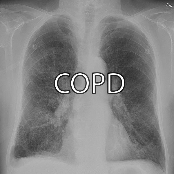
These can be fully compensated as you will see in chronic COPD, where pH is normal but the patient is in Type 2 respiratory failure - CO2 is high, the patient is hypoxic, but HCO3- retention compensates the acidosis.
In a respiratory alkalosis you see an increased pH and decreased PaCO2 due to increased ventilation rate e.g. hyperventilation.
In a metabolic acidosis you see decreased pH, decreased HCO3- and normal or low PaCO2 as ventilation rate quickly increases to compensate by blowing off CO2 (partial respiratory compensation).
Calculating the anion gap can help you narrow down the cause of a metabolic acidosis, using the equation (Na+ + K+) - (Cl- + HCO3-). The anion gap should be in the range 9-14 mmol/L.
The traditional mnemonic for the causes of a metabolic acidosis with raised anion gap is ‘MUDPILES’:
• Methanol (because it forms formic acid)
• Uraemia (kidney failure)
• Diabetic ketoacidosis (and alcoholic/starvation ketoacidosis) - check the ketones
• Propylene glycol
• Isoniazid/Iron overload
• Lactate - this is on the ABG, due to tissue hypoxia in a number of disorders
• Ethylene glycol
• Salicylates - from aspirin overdose
Normal anion gap metabolic acidosis can be remembered by ABCD - Addisons, Bicarbonate loss (gastrointestinal or renal including renal tubular acidosis), Chloride, Drugs (espeically acetazolamide)
In a metabolic alkalosis you see an increased pH and a normal or increased PaCO2 due to decreased ventilation rate (partial respiratory compensation). The alkalosis is due to acid loss or excess bicarbonate.
Common causes are direct loss of H+ from the stomach (vomiting, NG suction), loss from the kidneys in Conn's syndrome, hypokalaemia (causing shift of H+ into cells), or excess bicarbonate (antacids or IV)
Tip: In primary respiratory disorders, the pH and CO2 go in opposite directions, while in primary metabolic disorders they go in the same direction.
Base Excess:
Indicates metabolic acidosis or alkalosis:
High base excess (>+2 mmol/L): Possible metabolic alkalosis or compensated respiratory acidosis.
Low base excess (<-2 mmol/L): Suggests metabolic acidosis or compensated respiratory alkalosis.

You can buy the Reference Ranges YardCard with the ABG interpretation tool here!


Leave a comment